


Image: MomJunction Design Team
Different types of fetal ultrasound scans are performed to ensure the healthy progress of the pregnancy. The transvaginal scan during pregnancy is done by inserting a scanning instrument in the vagina. It helps doctors check the reproductive organs of a pregnant woman and assess the fetal health.
This procedure is simple and painless and is usually done during the early stages of pregnancy. However, your doctor may advise you to get it done later based on your pregnancy.
Read this post for detailed information on transvaginal ultrasound scans, including why and how it is done and how you can prepare for it.
What Is A Transvaginal Scan?
It is a safe and effective, noninvasive diagnostic scan that provides images of the female pelvic area including the bladder, ovaries, fallopian tubesiXTube-like structures connecting the ovaries and uterus that help transport eggs for the fertilization process. , cervix, and uterus. In pregnant women, the test helps to assess the pregnancy and health of the growing fetus. It can also help detect the placenta placement and cervical length and is used in amniocentesis. It measures various parts of the fetus such as the spine, head, and the heart (1) (2).

Image: Shutterstock
In some cases, a transvaginal scan may be recommended over an abdominal scan for detailed visualization of the fetus and fetal position. The test is done using a thin and long transducer, which is inserted into the vagina. This instrument is also known as a probe and is covered in latex or plastic sheath. A clear conducting gel is applied to it before the scan. The probe used in the ultrasound consists of a microphone, which sends high frequency sound waves (ultrasound) through the vagina into the pelvic cavity which are then reflected back to the microphone. These sound waves are converted to electrical impulses that produce a moving image on the computer screen..
Some other names of the transvaginal ultrasound include pelvic scan, gynecologic ultrasound, endovaginal ultrasound, transvaginal sonography, and pelvic sonography.
What Does A Transvaginal Ultrasound Show?
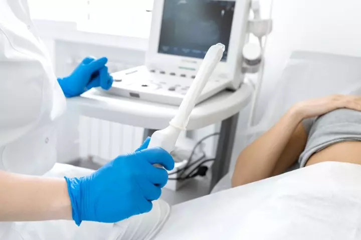
Image: Shutterstock
The ultrasound shows any changes that happen in the mother’s womb, the ovaries, or the organs surrounding it. In addition to this, it helps track fetal development and detect any problems related to fetal health so that they can be managed early on.
In rare cases, a tissue sample can be taken during the scan for further assessment. In addition to this, a hysterosalpingogram could also be done at the time of the ultrasound to check the inside of the uterus and patency of the fallopian tube (3) (4).
- Identify any issues with the endometrial liningiXA lining of the uterus where the embryo implants after the fertilization process. of the uterus
- Checks for pregnancy complications such as ectopic pregnancy
- Check if there are any infections or pelvic pain
- Monitor the fetus such as the fetal heart rate and identifies issues, if any
- Looks for cysts or fibroidiXBenign uterine growths that may cause heavy menstrual bleeding, contractions, and pelvic and back pain. or any abnormalities
- Diagnose high-risk pregnancies, any possible miscarriage, or preterm labor or premature delivery complications
 Quick fact
The main objective of TVS in pregnancy is to assess fetal health and to detect any complications. Based on these parameters, the doctor will determine the best time for a scan and how to do it. Find out more about it next.
When Is The Transvaginal Scan Done During Pregnancy?
It is usually recommended during the early stages of pregnancy, which is the first trimester or the beginning of the second trimester. Most women get this done between 10 and 14 weeks, while others could get it done earlier or at a later date (7) (8).
In any case, the doctor will determine when the ideal time for this scan is and can help you prepare for it.
Quick factHow To Prepare For A Transvaginal Ultrasound?
Considering that the scan is through the vagina, you could become anxious before the procedure. Keep reading to know how exactly the procedure works and how to prepare for it to bring down some of that relevant anxiety.
Here are a few points to remember before you go for the ultrasound (9) (10).
- The transvaginal ultrasound is a simple, risk-free, and painless procedure.
- Your sonographer will explain the procedure to you, so try to relax and understand the step-by-step process. Also, clear your doubts without hesitation.
- In case you are allergic to something, let the medical professional know about it.
- You would be asked to empty your bladder before the procedure.
- There will be a female nurse or attendant with you even if a male sonologist is performing the scan.

Image: Shutterstock
- The scan will be done by inserting the probe only with your consent, so you don’t have to worry about it.
You may even request to insert the probe yourself, like Annie, a mother who recalls her experience with a transvaginal scan. She says, “When I went in for my first endovaginal ultrasound, the tech let me guide the probe in myself, and it made a big difference. My second endovaginal ultrasound was performed by a midwife, and she placed the transducer herself — and it was much more uncomfortable. If you want to, go ahead and ask if you can guide the transducer yourself (i).”
Transvaginal Ultrasound Procedure
The procedure takes about 30 to 60 minutes. All you have to do is follow the instructions of the sonographer and stay relaxed (11).
- The sonographer might ask questions about your health, pregnancy, medications, symptoms, or surgeries.
- You might be asked to remove any jewelry or accessories that may interfere with the procedure. You will need to wear a hospital gown.
- You need to lie on the table with legs apart for the pelvic examination.
- A thin and long transvaginal transducer, covered in latex or plastic sheath, is lubricated to be inserted into your vagina.
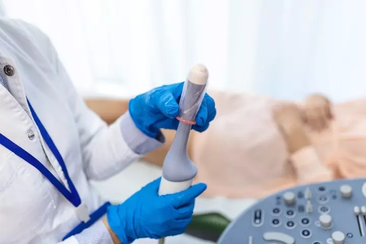
Image: Shutterstock
- Initially, you might feel some discomfort. The transducer could be slightly turned to get better images of your pelvic region.
- Once the images are recorded, the probe is removed.
Quick factAfter the procedure, you can get back to your regular diet and day-to-day activities. If the scan detects any abnormalities or issues, the doctor will give you additional guidelines.
The results of the scan can be collected at a date specified by the sonographer. It usually takes 24 hours or less. You can show it to your doctor for further assessment.
Although relatively safe, a transvaginal scan could have some risks. Find out about it next.

Image: Shutterstock
There are no major risks associated with the transvaginal ultrasound procedure. However, certain conditions such as severe obesity, filled bladder, sensitivity to latex, and gastro issues could interfere with the scan results. You can let your doctor know about them beforehand and fix it.
The only flip side to this scan could be any discomfort associated with it. If, at any time during the procedure, you experience pain, tell the sonographer about it.
Frequently Asked Questions
1. How many transvaginal ultrasounds can I have during pregnancy?
A transvaginal ultrasound is an internal ultrasound performed in the early stages of pregnancy by inserting a tubular probe into the vaginal canal (12). Your OB-GYN may decide on the number of times this ultrasound can be performed based on your overall health and the stage of pregnancy.
2. Is a transvaginal ultrasound necessary in early pregnancy?
A transvaginal ultrasound during early pregnancy helps determine the gestational age. It helps see changes in the womb, ovaries, and uterus (3) (12). Doctors may recommend this test to detect any problems in the initial stages of pregnancy and treat them to preempt any complications in the later stages and delivery.
3. Can a transvaginal scan detect the gender of the baby during pregnancy?
Transvaginal sonography can be used to determine the gender of a baby during early pregnancy. However, determining the gender too early, between 13 and 16 weeks, can be risky and inaccurate. During the early stage, the baby’s external genitalia look different from the later stages of pregnancy (14).
Your obstetric/gynecology specialist may recommend a transvaginal scan during pregnancy to check fetal health or identify any pregnancy complications during the early stages of the pregnancy. Your doctor will determine the right time to perform this scan. The procedure is non-invasive and usually pain-free. However, if you experience any discomfort, you can always inform the nurse. It may also help you discuss the procedure in advance with your health professional and soften your worries. After the test, your medical advisor would provide further guidelines based on their observation.
Infographic: Things You Should Know About A Transvaginal Ultrasound
Besides following your doctor’s advice, there are a few other essential points to learn about a transvaginal ultrasound before going for the test. So, check out this infographic as we present valuable tips to help you stay calm and cooperative during the procedure. Illustration: Momjunction Design Team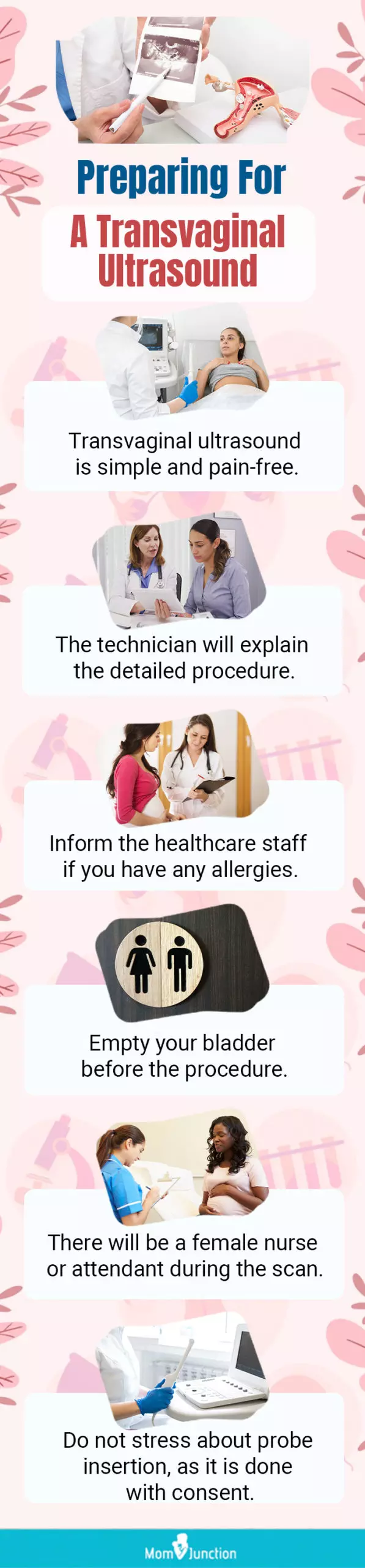


Key Pointers
- Transvaginal ultrasonography is a diagnostic test that produces images of a pregnant woman’s pelvic region.
- It is usually advised during the first or early second trimester of pregnancy, although the doctor decides the best to perform the test.
- Women are advised to empty their bladder before the procedure, wear a hospital gown, and follow the sonographer’s instructions.
- The procedure is simple, painless, and typically takes 30 to 60 minutes.
- The test helps assess the pregnancy and fetal health, and can detect issues such as ectopic pregnancy, infections, cysts, fibroids, or other abnormalities.
Ultrasounds are a gateway to visualize your little one throughout pregnancy. Explore the disparities between transvaginal and abdominal ultrasounds and pinpoint the ideal choice for your unique circumstances through this informative video.
Personal Experience: Source
MomJunction articles include first-hand experiences to provide you with better insights through real-life narratives. Here are the sources of personal accounts referenced in this article.
i. Your first transvaginal ultrasound: you’re going to put that where?;https://theshygirlsguide.wordpress.com/2008/10/25/your-first-transvaginal-ultrasound-you%E2%80%99re-going-to-put-that-where/
References
1. Fetal ultrasound; Stanford Children’s Health
2. Pelvic Ultrasound; Johns Hopkins Medicine
3. Transvaginal ultrasound scan; Cancer Research UK
4. Pelvic ultrasound; Kaiser Permanente
5. What is pelvic ultrasound; University of Utah Health
6. Ultrasound; USF Health; University of South Florida
7. First trimester screening; University of Rochester Medical Center
8. Is early second trimester vaginal ultrasound scan associated with adverse perinatal outcomes?; NIH U.S. National Library of Medicine (2009)
9. R. S. Moorthy; Transvaginal sonography; Medical Journal, Armed Forces India (2000)
10. Understanding pelvic ultrasound; Beaumont.org
11. Transvaginal ultrasound; NIH; U.S. National Library of Medicine
12. Pregnancy: Prenatal Ultrasonography; Cleveland Clinic
13. Vaginal Ultrasound; Pregnancy, Birth and Baby
14. Early determination of fetal sex using transvaginal sonography: technique and pitfalls; NIH


 For Babies Is It Safe, Tips And Alternatives")


