
Image: Shutterstock
Lennart Nilsoon, a Swedish photographer has devoted twelve years of his life capturing images of foetus developing in the womb. The stunning photographes were taken using conventional cameras with macro lenses, a scanning electron microscope and an endoscope. His first image of the human foetus was taken in 1965. Nilsson had to employ magnification by hundreds of thousands to capture the photos right from the womb. Advanced technology now allows even clearer images.
Image 1
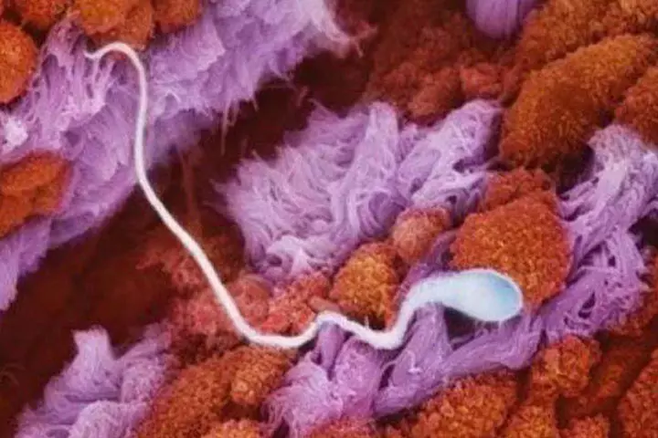
This image shows the sperm in fallopian tube.
Image 2
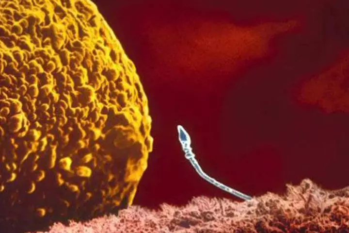
It’s a moment of suspense that keeps you guessing if the sperm will fertilize the egg.
Image 3
Two sperms jostling to get the egg cell.
Image 4
One of them wins.
Image 5
The human embryo attaches to the uterine wall.
Image 6
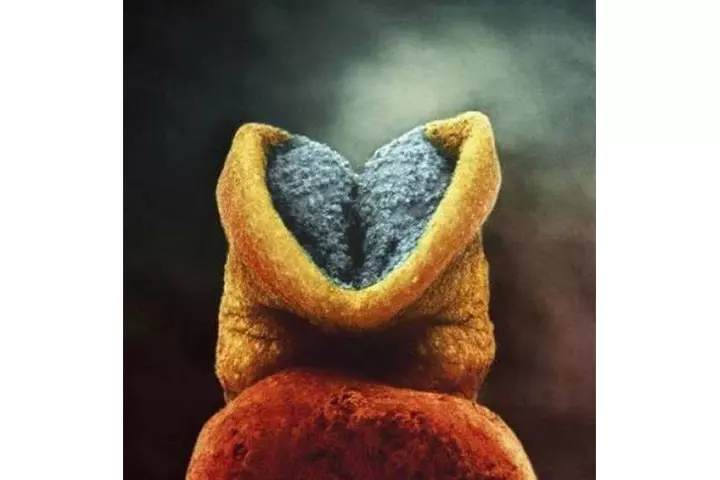
The brain begins to develop. The child begins to feel everything already.
Image 7
Image of a 24 day embroy. The heart starts beating on the 18th day.
Image 8
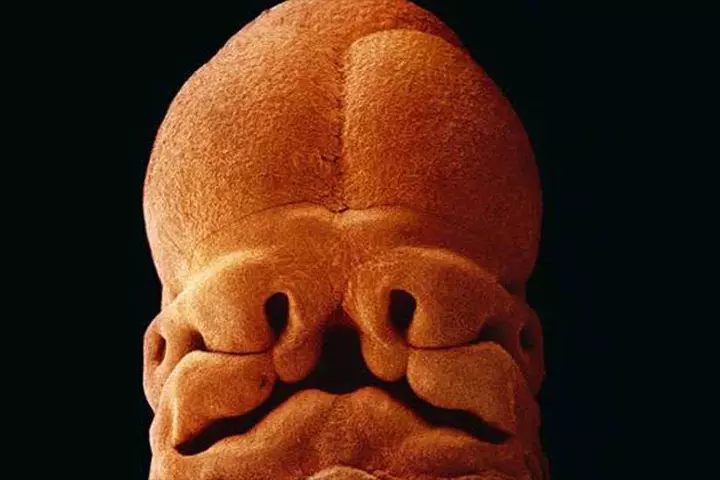
At 5 weeks, you can see the eyes, the nostril and the mouth developing.
Image 9
At 8 weeks
Image 10
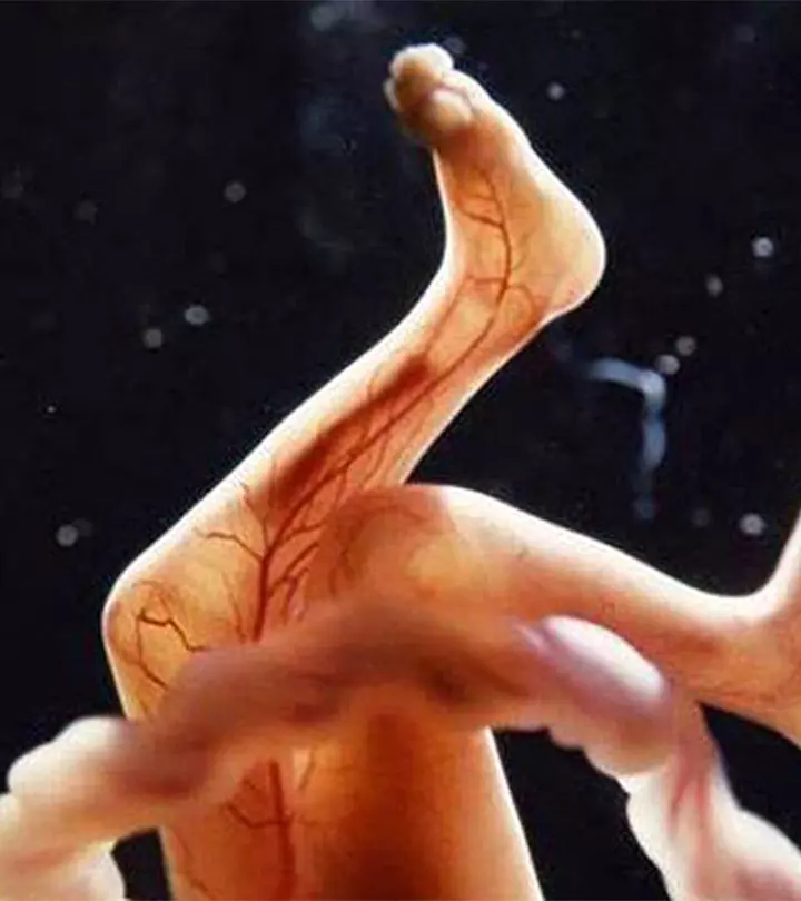
You can see the blood vessels.
Image 11
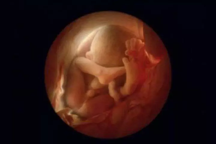
At 18 weeks, it is about 14cm. It is receptive to sounds from outside the womb.
Image 12
The baby grows nails.
Image 13
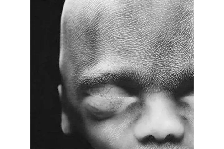
The foetus measures 24 cms by now. The image shows the forehead being covered with lugano – woolly hair.
Image 14
At 26 weeks, the baby’s eyes are still developing. It could respond to increased activity through increased pulse rate.
Image 15
At 36 weeks.
Community Experiences
Join the conversation and become a part of our nurturing community! Share your stories, experiences, and insights to connect with fellow parents.

