Image: Shutterstock
The 5th-week ultrasound can help detect any irregularities in the pregnancy. It gives the parents the first glimpse of their developing baby. An ultrasound may be prescribed around this stage to identify any possible complications. It is better to wait for at least 12 weeks of pregnancy for an otherwise healthy mother.

Read this post to understand the importance of undergoing an ultrasound during week 5 of pregnancy care.
Key Pointers
- Five-week ultrasound scan is a part of regular antenatal care for women who have a history of miscarriage or have conceived via IVF.
- Vaginal bleeding after pregnancy confirmation might also need this scan.
- This endovaginal scan helps determine embryo growth, gestational age, and embryo’s heart rate.
- If the doctor observes any issues in the yolk sac, gestational sac, or fetal pole, they will decide the course ahead accordingly.
When To Go For The First Ultrasound Scan?

According to the American College of Obstetricians and Gynecologists, a woman can go for a pregnancy ultrasound scan between 18 and 22 weeks of pregnancy (1). However, each pregnancy is different, and the doctor will decide the first and subsequent ultrasound visit.
Why Is 5-Week Ultrasound Important?
The week five ultrasound scan is part of regular pregnancy care, and it should usually show developmental milestones. While experts generally recommend you visit a midwife or a doctor at around six to eight weeks of pregnancy (2), an early ultrasound at five weeks can reassure expectant parents by confirming the presence of the gestational sac and embryo.
An endo-vaginal ultrasound scan is essential to detect the following (1) (3).
- Growth of the embryo
- Gestational age
- Due date
- Genetic disorders
- Multiple pregnancies
- Embryo’s heart rate
- Ectopic pregnancies
Generally, a 5-week ultrasound should show developmental milestones.
According to peer-reviewed studies, if nothing is seen in the ultrasound, it could mean the following (4).
- Pregnancy might be too early to show up in the ultrasound scan
- Ectopic pregnancy, in which the embryo implants outside the uterus. In this case, the embryo will not survive, and it can often lead to severe complications for the mother.
- Pregnancy might have gone through a miscarriage.
 Quick fact
Sharing her experience from her fifth-week ultrasound scan, Savannah Glembin, a mom, says, “I’m just super nervous because I don’t really know what to expect. Even though I am on my medications because of my blood disorder, it’s just hard for me to want to love my little being because I’m scared that I might miscarry. Observed the gestational sac with the help of my tech and just hoped that the baby was healthy (ⅰ).’’
The ultrasound is a non-invasive procedure, and it does not affect pregnancy. However, the overuse of ultrasound on the developing embryo can have unknown harmful effects.
What Are The Precautions For A 5-Week Ultrasound?
Some of the precautions required for a 5-week ultrasound scan are (5) (6):
- There is no scientific evidence regarding the harmful impact of ultrasound on pregnancy. However, some studies suggest avoiding excessive and unnecessary ultrasound exposure as it may risk the developing embryo.
- 3D and 4D sonograms are unnecessary unless in special medically required cases.
- Sometimes, more ultrasounds may be done within the trimester if there are problems or your pregnancy is considered high-risk.
What Can You See On A 5-Week Ultrasound?
You can expect the following diagnosis during an ultrasound scan when 5 weeks pregnant (7) (8) (9):
- Location of the embryo that looks curled and is around 2 mm in size.
- Gestational sac, a fluid-filled structure around an embryo, will be visible beyond 4.5 weeks. It gives a 97.6% confirmation for an intrauterine pregnancy (IUP).
- Yolk sac, a circular structure seen within a gestational sac, becomes visible in 5.5 weeks. Visibility of the yolk sac is a 100% confirmation for an IUP.
- In normal pregnancies, a beating heart might be visible. However, parents should not get worried if they cannot see a beating heart at this stage. It is entirely normal as every pregnancy is unique.
 Research finds
If the imaging test detects any abnormalities in the fetal pole, yolk sac, and gestational sac, the healthcare provider will address the condition based on the severity.
How To Prepare For A 5-Week Ultrasound Scan?
Some of the vital preparations required for a 5-week ultrasound exam are (10):
- You should have a full bladder. Drink water and do not go to the toilet until after the scan.

- You can have regular food before the fetal ultrasound.
- Wear comfortable clothing that you can easily remove to facilitate vaginal examination using an ultrasound probe.
- Present the list of your current medications to your doctor to ensure they are safe for your pregnancy.
How Is A 5-Week Ultrasound Performed?
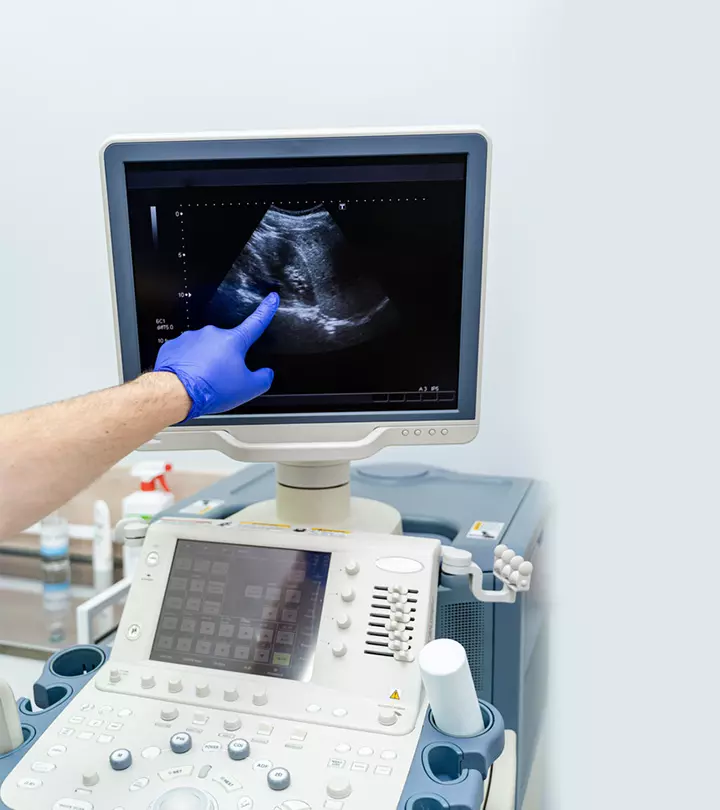
The World Health Organization (WHO) mandates at least one ultrasound scan throughout pregnancy (11). The ultrasound scan gives an accurate estimation of the size of the embryo and the delivery date. In the 5th week of gestation, ultrasound is carried out in the transvaginal route instead of the abdominal.
- A small transducer covered with a sterile lubricated condom is inserted into the vagina.
- The transducer transmits sound waves and records the reflections.
- The ultrasound machine interprets the echoes in the form of an image or video, which are displayed on a screen
Though not painful, this procedure could cause some discomfort.
How Long Does A 5-Week Ultrasound Take?

In general, the whole ultrasound procedure takes around 30 minutes to one hour (12). If another patient is in line, it might add the waiting time to the procedure.
Can A 5-Week Scan Detect Twins?
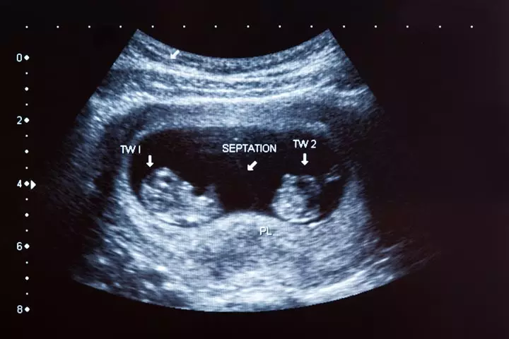
Ultrasound can detect twins between six and eight weeks. However, a 5-week ultrasound is too early to detect if twins are present in the same sac. A doctor might determine the patterns of fetal heartbeat to detect twins; however, there is a chance to miss this.
Ultrasounds conducted during pregnancy help doctors regularly monitor the growth and development of the fetus. The fifth-week ultrasound is conducted to ensure the developing baby has no genetic anomalies. Hence, you should visit your doctor regularly for checkups and ultrasounds to ensure there are no probable complications. Further, if any anomalies are detected during these visits, they may be effectively treated with early intervention. Take proper care of yourself from the beginning of the pregnancy, and consult your gynecologist if you have any questions or concerns.
Frequently Asked Questions
1. Can I see a miscarriage at five weeks on ultrasound?
A five-week ultrasound may not accurately show a miscarriage. It may be difficult to identify a heartbeat early in pregnancy, and whether a fetus has died, failed to develop, or is smaller than expected may not be fully understood (13).
2. What is the size of the gestational sac as seen on the fifth-week ultrasound scan?
At around five weeks of pregnancy, the gestational sac appears centrally in the uterus and measures two to three millimeters in diameter (14).
3. How soon after a 5th week ultrasound will I receive the results?
The scan itself may be for around half an hour and you may be told the results of your ultrasound soon after your scan is over but the images are usually assessed, analyzed and forwarded to the doctor that referred you to get the scan done. The results of which will be revealed and explained at the next consultation (15).
Infographic: Key Findings At Your 5-Week Pregnancy Ultrasound
If you have a high-risk pregnancy, an early 5-week pregnancy ultrasound can give you some insights into your unborn baby and its health. The scan may detect any possible complications or anomalies your fetus might be facing. Look through the infographic below to know what you may expect at your 5-week ultrasound during pregnancy.
Some thing wrong with infographic shortcode. please verify shortcode syntaxIllustration: What You&rsquoll See At 5-Week Ultrasound & How To Prepare For It

Image: Stable Diffusion/MomJunction Design Team
Personal Experience: Source
MomJunction articles include first-hand experiences to provide you with better insights through real-life narratives. Here are the sources of personal accounts referenced in this article.
ⅰ. My scary first ultrasound (5 weeks 5 days) // Pregnancy vlog 3.https://www.youtube.com/watch?v=2r-Ssx5twdA
References
- Ultrasound Exams.
https://www.acog.org/womens-health/faqs/ultrasound-exams - Your first antenatal visit
https://www.pregnancybirthbaby.org.au/your-first-antenatal-visit - Early pregnancy ultrasound measurements and prediction of first trimester pregnancy loss: A logistic model.
https://idp.nature.com/authorize?response_type=cookie&client_id=grover&redirect_uri=https%3A%2F%2Fwww.nature.com%2Farticles%2Fs41598-020-58114-3 - The Use of Ultrasonography in the Diagnosis of Ectopic Pregnancy: A Case Report and Review of the Literature.
https://www.ncbi.nlm.nih.gov/pmc/articles/PMC2270872/ - The accuracy of ultrasound estimation of fetal weight in comparison to birth weight: A systematic review.
https://www.ncbi.nlm.nih.gov/pmc/articles/PMC5810856/ - Safety of ultrasonography in pregnancy: WHO systematic review of the literature and meta-analysis.
https://www.ncbi.nlm.nih.gov/pmc/articles/PMC5810856/ - Gestational Sac Evaluation.
https://www.ncbi.nlm.nih.gov/books/NBK551624/ - What will I see at a 5 week early pregnancy scan, why should I wait until 7 weeks?
https://www.earlylife.co.uk/blogs/news/what-will-i-see-at-a-5-week-early-pregnancy-scan-why-should-i-wait-until-7-weeks - Pregnancy at week 5.
https://www.pregnancybirthbaby.org.au/pregnancy-at-week-5 - How To Prepare for an Ultrasound During Pregnancy.
https://www.beaumont.org/treatments/ultrasound-how-to-prepare - WHO recommendations on antenatal care for a positive pregnancy experience.
https://iris.who.int/bitstream/handle/10665/250796/9789241549912-eng.pdf;jsessionid=271288604455CCAF852818566A7FE0AF?sequence=1 - Transvaginal Ultrasound.
https://www.cedars-sinai.org/programs/imaging-center/exams/ultrasound/transvaginal.html - Ultrasound scans.
https://www.miscarriageassociation.org.uk/information/worried-about-pregnancy-loss/ultrasound-scans/ - Olga Dewald and Jennifer T. Hoffman; (2025);Gestational Sac Evaluation.
https://www.ncbi.nlm.nih.gov/books/NBK551624/ - Ultrasound Scan.
https://www.nhs.uk/conditions/ultrasound-scan/#:~:text=You%20may%20be%20told%20the,appointment%2C%20if%20one’s%20been%20arranged.
Community Experiences
Join the conversation and become a part of our nurturing community! Share your stories, experiences, and insights to connect with fellow parents.
Read full bio of Dr. Richa Hatila Singh
Read full bio of Anshuman Mohapatra
Read full bio of Rebecca Malachi
Read full bio of Aneesha Amonz


