Image: Shutterstock
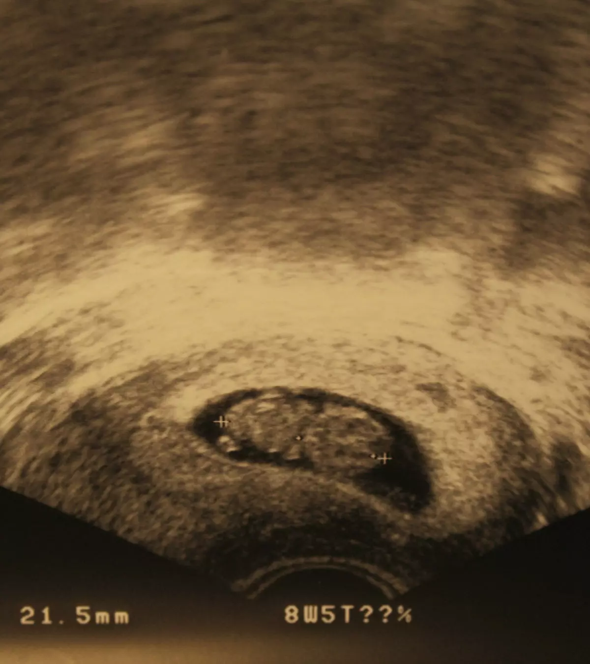
The 8th-week ultrasound is usually the first imaging scan in your pregnancy. It is very exciting for the parents as this scan gives you the first glimpse of your baby. This scan is also called the dating scan. It uses sound waves to create a picture of the fetus in the womb, called a sonogram.

Read this post as we tell you about the ultrasound procedure, what you can see at eight weeks of pregnancy, the fetus’ development, and what abnormalities may be discovered in the eight-week of pregnancy scan.
Key Pointers
- The 8th-week ultrasound helps confirm pregnancy, identify multiple gestations, and check the embryo size.
- You are supposed to keep your bladder full for an abdominal ultrasound and empty it if it is a transvaginal ultrasound.
- If the ultrasound fails to detect a heartbeat in the 8th week, the doctor may suggest a follow-up scan to look for it.
Why Is The 8th-Week Scan Done?
The first ultrasound is usually done around the eighth week to monitor whether both the expectant mother and the growing baby are fine.
It might also be suggested to (1) (2):
- Calculate the gestational age and due date. It is also done when you are not sure about your last period date (which can give an approximate gestational age)
- Know the cause of bleeding, if any, in the pregnant mom
- Check the embryo size and the heartbeat
- Confirm multiple pregnancies
- Check if the ovaries and fallopian tubes are healthy
- Rule out pregnancy complications such as ectopic pregnancyiA condition where a fertilized egg implants and develops outside of the uterus, such as in the fallopian tube , miscarriage
How To Prepare For Your 8th-week Scan?

Your doctor can recommend either the abdominal ultrasound or the transvaginal ultrasound (3) (10) (12).
- For the abdominal scan, you will be asked to have a full bladder. At this stage of pregnancy, the fetus is tiny, and a full bladder helps move the uterus up to get a better image of the fetus. However, to prevent the bladder from being too full, empty it 90 minutes before the scan and consume eight ounces of water an hour before the test. Wear comfortable and loose clothing since you will have to expose your abdomen during the scan.
- For the transvaginal scan, you will be asked to empty your bladder since a full bladder blocks the fetal view.
The Ultrasound Procedure
A sonographer performs the pregnancy ultrasound scan, which is neither harmful nor painful for the mother and the baby (1).
- Abdominal ultrasound scan: A conductive gel is applied over the abdomen, and a handheld scanner is run over it. This gel allows the ultrasound waves to pass into the uterus through the skin. The waves will then rebound, creating a picture of the fetus in the uterus. Abdominal ultrasounds are available in two, three, or four dimensions and include the Doppler scan and the fetal echocardiogram.
- Transvaginal ultrasound scan: Here, a small wand called the transducer is placed inside the vagina and pressed against the cervix. It may be moved around a little for the right angle to view and capture an image of the pelvic organs and the fetus. You may experience a little pressure or discomfort during the procedure.

What Is The Duration Of The Ultrasound?
The first obstetric ultrasonography takes up to 30 minutes (4). In some cases, it might take longer if the baby is positioned unusually, or if the body tissues are dense.
What Can You See At 8th Week Ultrasound?
The 8th-week ultrasound examination can give you a great deal of information including the (5):
- Umbilical cord functioning
- Size of the placenta
- Size of the embryo (which may be about 1/4 to 1/2 inch at eight weeks)
- Fetal heartbeat and heart rate
- Presence of multiple fetuses that can be detected through the presence of multiple heartbeats
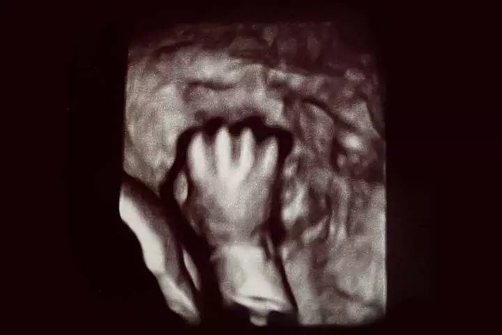
- The growth of tiny hands, legs, the formation of internal organs, eyes, nostrils, and mouth
 Did you know?
How Is The Baby Developing In The 8th Week?
The baby will attain basic human features and is now called a fetus, but not an embryo (13).
- The baby has tiny webbed arms, legs, with slight bends where the elbows and knees will develop
- Face and internal organs (such as heart, liver, and sex organs) begin to take shape
- A tiny mouth, upper lip, nostrils, eyes, eyelids, and tooth buds begin to form
- The baby begins to move, but you may not feel it (2)
- Heart rate would be around 140 – 160bpm
A rhythmic heartbeat can be detected using sonography around the 6th week of gestation. The graph below demonstrates the changes in fetal heart rate through gestation.
According to the findings, the fetal heart rate elevates from 110 beats to 170 beats during the gestational period of six to nine or ten weeks. However, at approximately 14 weeks, the heart rate decreases to roughly 150 beats and ultimately lowers to about 130 around the term.

Fetal heart rate in the first trimester
Source: Fetal heart rate in the first and second trimester; Radiopedia Quick fact
If the Ultrasound Doesn’t Detect The Fetal heartbeat, Does It Indicate A Miscarriage?
No, it does not. If the heartbeat is not detected in the eight-week ultrasound, your doctor will suggest a follow-up scan to check for the pulse again and rule out the possibility of a miscarriage.
What If Any Other Abnormalities Are Found In The 8th Week Pregnancy Scan?
If the ultrasound shows any abnormalities in the 8th week pregnancy scan, the sonographer or medical professional might suggest another scan.
In some cases, they might suggest diagnostic tests such as amniocentesisiA prenatal diagnostic test in which a sample of amniotic fluid is withdrawn from the uterus to analyze fetal cells for genetic defects and other conditions. or CVSiAlso known as chorionic villus sampling, this prenatal test is to diagnose chromosomal or genetic disorders by testing the cells from the placenta or the chorionic villi to detect the problem.
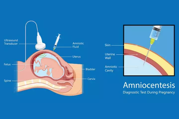
The 8th-week ultrasound happens to be considered a little early. It so happens that in most cases, doctors forgo it as it may not be necessary. However, you may still have an ultrasound to know the due date by understanding the gestational age. Also, you must note that the ultrasound reports may differ from case to case. You need not panic seeing your results as it is too early to detect the things. Lastly, be sure to talk to your doctor to avoid doubts and fears about the pregnancy.
Frequently Asked Questions
1. Can an 8th week ultrasound miss twins?
It is possible to miss twin pregnancies on the 8th-week ultrasound. However, there is less chance of missing twin pregnancy since two embryos with fetal heart rate can be found by this time. Usually, a sonographer can visualize two gestational sacs in the uterus from six weeks onwards (6).
2. Why do doctors wait until eight weeks for an ultrasound?
If the first ultrasound scan in six weeks does not show a developing fetus with a heartbeat, doctors may schedule the next ultrasound for the 8th week. Even if you do an ultrasound on the 7th week, you may have to do an ultrasound on the 8th week for clear visualization. To avoid multiple visits, doctors may ask you to wait until the 8th week for the next ultrasound (7).
3. Can we determine the gender of the baby during an 8th-week ultrasound?
The fetal gender is easily detectable around weeks 18 to 20 of pregnancy, however, in some countries, its revelation may be prohibited by doctors (10).
4. How does the size and appearance of the fetus change from week 8 to week 9?
From week eight to nine, the size and appearance of the fetus change as its arms and legs develop joints and fingers. Additionally, the internal organs and facial features continue to develop, and the formation of nipples and hair follicles begins (11).
5. What should I ask my doctor before the ultrasound?
Before your ultrasound, you may ask your doctor about the type of ultrasound, its purpose, the duration, whether you can see the images, when you can expect the results, any necessary preparations, and how to address any post-ultrasound concerns.
Infographic: Which Is Better, Transvaginal Ultrasound Or Transabdominal Ultrasound In 8th Week?
Prenatal ultrasound is recommended at specific intervals to observe the growing baby and placenta. Doctors may recommend the type of ultrasound based on the required image and other factors. Go through the infographic to learn more about transvaginal and transabdominal ultrasound during pregnancy.
Some thing wrong with infographic shortcode. please verify shortcode syntax
During the eighth week, your baby is growing rapidly, and you might experience some noticeable changes in your body.
References
1. Ultrasound In Pregnancy; Stanford Children’s Health
2. Normal Pregnancy; Operational Obstetrics & Gynecology – 2nd Edition (2000)
3. What Is A Pelvic Ultrasound; University of Utah Health
4. The Biophysical Profile; Texas Tech University Health Sciences Center El Paso Department of Obstetrics and Gynecology
5. O. Valenti et al.; Fetal cardiac function during the first trimester of pregnancy; J Prenat Med (2011)
6. S Petaković, et al; [Abnormal twin pregnancy–early resorption of a single fetus in a twin pregnancy pregnancy]; National Library Of Medicine
7. Patience is key: Understanding the timing of early ultrasounds; UT Southwestern Medical Center
8. Early Pregnancy Scan/Viability Scan; Diagnostic Ultrasound
9. 8 weeks pregnant; Raising Children Network (Australia)
10. Ultrasound in Pregnancy; Cleveland Clinic
11. Fetal development; Mount sinai
12. How To Prepare for an Ultrasound During Pregnancy; Corewell Health
13. What happens in the second month of pregnancy?; Planned Parenthood Federation of America Inc.
Community Experiences
Join the conversation and become a part of our nurturing community! Share your stories, experiences, and insights to connect with fellow parents.
Read full bio of Lucy Mendelssohn
Read full bio of Rebecca Malachi
Read full bio of Dr. Ritika Shah
Read full bio of Aneesha Amonz


