Image: ShutterStock
When you think of pregnancy, the first thing you may envision is a growing belly. While this is a visible change of pregnancy that is hard to ignore, myriad transformations occur inside a pregnant woman’s body. The most important change is the size of the uterus during pregnancy.

The uterus serves as the abode of your baby and plays a crucial role in pregnancy, and it constantly expands during your gestation journey to hold the developing fetus.
This post discusses interesting details about the uterus, including its size during and before pregnancy, functions, and positions. It also mentions how to maintain a healthy uterus.
Key Pointers
- The uterus is your baby’s home, constantly expanding during the gestation period to hold the developing fetus.
- While pregnant, women may experience a constant need to urinate. This occurs because the ever-growing uterus puts pressure on the bladder.
- Involution or shrinking of the uterus occurs post-childbirth. It takes about six to eight weeks for the uterus to shrink back to its normal position and size, after which it starts preparing for the next menstrual cycle.
- Maintain a healthy uterus by eating the right foods, exercising regularly, consuming omega-3 fatty acid-rich food, quitting smoking, and not holding your urine.
Uterus During Pregnancy
The uterus is a distensible organ the size of a closed fist. It grows and changes to become large enough to accommodate a full-term baby. It is held in its position by ligaments, which stretch as the uterus grows.
Dr. Alan Lindemann, MD, a former clinical associate professor at the University of North Dakota, says, “When you get 10 or 12 weeks along, about the end of the third month, your uterus could be appreciably enlarged and feel heavy. You also have more blood supply, so your arteries and veins to and from your uterus might be perceived as heavy.”
Read on as we tell you about what exactly the uterus does, how much its size changes through the stages of pregnancy, and how you can measure the organ and keep it healthy.
Size Of Uterus During Pregnancy
As you know, the uterus keeps changing in shape and size as your pregnancy progresses. The uterus expands between 500 and 1,000 times its normal size (1). Let’s see how the organ changes during each trimester.
The first trimester (0 to 12 weeks)
- At 12 weeks of pregnancy, the size of the uterus remains as small as the size of a grapefruit.
- As your pregnancy progresses, the uterus grows, putting pressure on the bladder. This is when the frequency of urination increases (2).
- In the case of twins or multiple pregnancies, the stretching of the uterus will be faster compared to that of a single baby.
The second trimester (14-28 weeks)
- By the second trimester, the uterus grows to the size of a papaya. The uterus grows upward and develops outside the pelvic area.
- The uterus expands between the navel area and the breasts and starts putting thrust on the other organs, pushing them away from their original positions (3). This may lead to some tensions in the ligamentsiFibrous connective tissue that holds bones together to form a stable structure. and the surrounding muscles, leading to body aches and pains.
- In some cases, the naval may pop out because of the pressure put by the uterus on other organs.
The third trimester (28-40 weeks)
- By the third trimester, your uterus will get enlarged to the size of a watermelon. It will move from your pubic brim to the lower bottom of the rib cage.
- Once you approach labor, your baby will descend into the pelvis.
After childbirth
Postpartum, the uterus shrinks back to its normal position and size.
This process is known as involution, which will take about six to eight weeks (4).
Apart from changing in size to accommodate the growing fetus, the uterus also plays other roles during pregnancy.
Functions Of The Uterus During Pregnancy
During pregnancy, the uterus:
- Accepts the fertilized ovum that passes through the fallopian tubeiPair of tubes that carry eggs from ovaries to the uterus in women. .
- Creates the placentaiTemporary organ connecting the uterus to the umbilical cord, providing nutrients and oxygen to the fetus. for the development of the embryo and fetus.
- Nurtures the fetus (in the amniotic sac) with nutrients by developing blood vessels exclusively for this purpose.
- Contracts to facilitate the exit of the baby and the placenta through the birth canal (vagina) during childbirth.
- Post-delivery, it shrinks back and starts preparing for the next menstrual cycle.
- It helps the blood flow into the ovaries. It supports other organs such as the vagina, bladder, and rectum. It plays an important role in sexual functions like triggering orgasm (5).
During active labor, the uterus contracts in powerful, rhythmic waves to open the cervixiThe lowest part of the uterus connected to the vagina (dilation) and guide the baby through the birth canal. These contractions start at the top of the uterus and move downward, gradually becoming stronger, more regular, and more frequent (6).
Interesting how important this inconspicuous organ is, right? Read on, and we will give you more information about the uterus and its functions.
How Big Is A Uterus? Normal Size Of A Uterus In Women
The size of the uterus varies from woman to woman. It weighs approximately 70 to 125gm (7). However, the size of the uterus is based on factors such as age and hormonal conditions of a woman.
Size of the uterus:
- Before attaining puberty, the length of the uterus is about 3.5cm and thickness is approximately 1.4cm (8).
- After attaining puberty, the length is between 5 and 8cm, width is 3.5cm, and thickness is between 1.5 and 3cm.
- During pregnancy at term (between the 37th and 40th week pregnancy), the uterus measures 38cm in length and 24 to 26cm in width.
Just like the size of the uterus, the position of the uterus also varies from woman to woman. It can be in an anteverted, anteflexed, or any other position.
What Is The Uterus Made Of?
The uterus is an inverted, pear-shaped, and hollow muscular reproductive organ located in the pelvic region of a woman’s body (9).
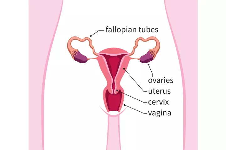
It is made of smooth muscles lined with various glands. These muscles contract during orgasm, menstruation, and labor, whereas the glands grow thicker with the stimulation of ovarian hormones every month.
The uterine wall consists of three key layers (10):
- Perimetrium
This is the outer layer of the uterus. It acts as a protective covering and helps keep the uterus in place within the pelvis. Additionally, a layer of fatty and fibrous connective tissue called the parametrium covers the outside surface and connects the uterine tissue to other pelvic tissues.
- Myometrium
The myometrium is the middle layer comprising smooth muscle fibers. It allows the uterus to stretch as the baby grows and contracts during labor to help with delivery.
- Endometrium
The endometrium, the innermost layer, has a mucus membrane. Each month, it thickens in preparation for pregnancy, with the uterine glands and blood vessels increasing in size and number. If a fertilized egg attaches to it, it supports the embryo. If not, it sheds during menstruation.
During pregnancy, these layers work together to support the baby. The perimetrium protects, the myometrium stretches and contracts, and the endometrium nourishes the baby. As the pregnancy progresses, the uterus expands, and each layer adapts to support the growing baby.
The uterus extends into the vagina through the cervix, which is made up of fibromuscular tissues, and it controls the flow of material into and out of the uterus.
 Did you know?
Positions Of The Uterus
One of the factors that influence the position of the uterus is the degree of dilation of the bladder. The uterus normally lies right above the bladder and in front of the (anterior) rectum. The normal uterine position is straight and vertical.
Common uterus positions
The uterus position can also be anteverted or anteflexed, both of which are common.
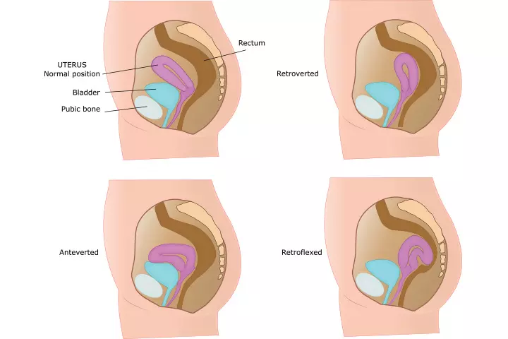
- In the anteverted position, the uterus is bent forward towards the bladder/ pubic bone and towards the front of the body.
- In an anteflexed position, the uterus is rotated towards the pubic bone with a concave surface.
Uncommon uterus positions
Certain positions are tilted back towards the rectum.
- A retroverted uterus is tipped backward toward the rectum. Around 25% of women have this type of uterus.
- A retroflexed uterus is the opposite of the anteflexed uterus, as it takes a concave shape towards the rectum (11).
A retro uterus is not known to create any fertility issues. However, you might have pain during sexual intercourse.
A tilted uterus during pregnancy usually does not cause problems, as the expanding uterus retains the forward-tipped position (12).
Measuring The Uterus During Pregnancy
Your doctor might measure the size of your uterus, also called the fundal height (fundus is the domed region at the top of the uterus), to understand the fetal growth and development. The fundal height is the measurement of the top of the pubic bone to the top of the uterus, which determines the gestational ageiTerm used to describe the duration of pregnancy in weeks. .
Katy, a mother of three from Colorado, shares her pregnancy update, “I had my 38-week appointment, and I was checked for dilatation. I was 1cm dilated at that appointment. My fundal height measured 37cm, which was the same fundal height that my belly has measured since about 34 weeks. She (the midwife) said breech babies cause unreliable fundal heights due to their positioning. So that makes sense that he (the baby) was making my belly measure unreliably bigger when he was previously in breech presentation. (At) my 39-week appointment my fundal height was 38cm (i).”
Note: It should be noted that the size of the uterus varies from woman to woman and depends on height, weight, and age (13).
You, too, can measure the size of your uterus. Before we go into the various methods of measuring it, let’s see the initial steps (14)
- Lie down on your back on the bed or the floor. While lying down, you should not feel any pain or dizziness.
- Locate your uterus by touching your tummy. If you are over 20 weeks pregnant, you can feel the uterus just above the navel. The uterus is hard round.
- Now, move your fingers upward to feel the top of the uterus, which is also referred to as the fundus.
- Proceed to locate your pubic bone. It is positioned just above your pubic hair line. The bone tip that you can feel is the pubic bone.
Once you locate the fundus and the pubic bone, you can measure the fundal height using various methods.
1. Measuring fundal height using a tape measure

- Measure the distance between your fundus and the pubic bone in centimeters using a tape.
- For example, if you are 24 weeks pregnant, then your uterus would usually measure 22-26cm. The uterus usually grows 1cm in a week or about 4cm in a month.
You can use this method to track your pregnancy growth week by week. However, studies say that more research needs to be done to find out the reliability of this method.
2. Measuring the fundal height using the finger method
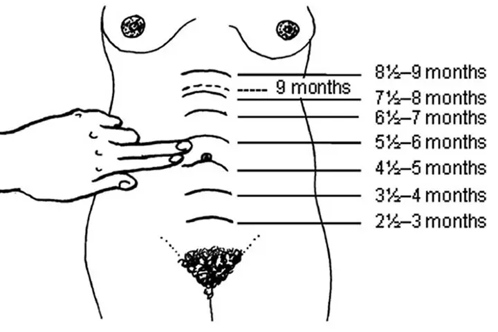
- If your uterus is below or above the belly button, then measure how many fingers below or above is the uterus from the belly button.
- The assumption is the fundal height increases by two finger-widths every month.
- For example, if you are 4 two-finger-widths above the belly button, then it indicates seven months gestation (see the above figure).
This method helps determine the gestational age in months (14).
If you find the fundal height different from the gestational week that you think you are in, talk to your doctor, and get your doubts cleared.
The size of the uterus during pregnancy plays an important role in the well-being of your baby growing inside.
Two uterine conditions that can affect pregnancy are fibroids and endometriosis (15) (16):
1. Uterine fibroids
Fibroids are harmless growths in the uterus. The American Academy of Family Physicians indicates that the prevalence of uterine fibroids is strongly linked to age, with up to 80% of women being diagnosed by the age of 50 (17). Uterine fibroids might:
- Increase miscarriage risk, especially in early pregnancy.
- Cause early labor.
- Cause placental problems in rare cases.
2. Endometriosis
It occurs when uterine-like tissue grows outside the uterus. The World Health Organization (WHO) estimates that about 10% of women and girls of reproductive age may have endometriosis worldwide (18). It can:
- Increase the risk of miscarriage.
- Raise the risk of placenta previa.
- Lead to postpartum hemorrhageiExcessive bleeding following childbirth .
- Cause uterine rupture.
Women with these conditions should work closely with their doctors during pregnancy for the best care. Next, we look at how you can maintain the health of your uterus during pregnancy.
How To Keep Your Uterus Healthy?
The best way to keep your uterus healthy is to keep yourself healthy. Here is how you can do it:
- Make sure to eat right and engage in some forms of exercise to maintain.
- Eat omega-3 fatty acid-rich food.
- Quit smoking as it has harmful effects on the uterus.
- Do not hold your urine.
Frequently Asked Questions
1. When does the mouth of the uterus open in pregnancy?
The opening of the mouth of the uterus is called cervical dilation. The cervix begins to dilate towards the end of pregnancy. It becomes thinner and dilates during the first stage of labor or uterine contractions, and is completely dilated by about 10cm during the last stage of labor (19).
2. How does a weak uterus affect pregnancy?
An incompetent or weak cervix, a kind of womb abnormality, may lead to preterm birth in some women (20).
3. Does a swollen uterus mean I am pregnant?
“A swollen uterus could mean pregnancy. Generally speaking, by the time you can appreciate the swelling in your uterus, you will be about three months along. However, there are other reasons for swelling. The most common reason for swelling is fibroids, which are benign smooth muscle tumors,” observes Dr. Lindemann.
4. Why do I feel a sharp pain in my uterus while pregnant?
Pregnant women may feel a stabbing pain in and around the uterus, stomach, or groin area. Cramping (as the uterus expands), round ligament pain (as the ligament that supports the uterus stretches with progressing pregnancy), gas, or constipation are some of its common causes (21).
5. Where is uterus pain located?
Uterus pain in early pregnancy is mostly felt in the pelvic floor area (22).
6. What are the signs of a healthy uterus during pregnancy?
A healthy uterus during pregnancy typically shows consistent growth as the baby develops. You should experience no severe pain, though mild discomfort can be expected. Regular fetal movement is a positive sign. Ultrasounds should show normal amniotic fluid levels. These signs generally indicate that the uterus is providing a safe, nurturing environment for the growing fetus (6).
The uterus expands during pregnancy, and its growth indicates the size of the fetus. After birth, the uterus again conforms to its previous size. It is beautiful how a small organ grows over time into the size of a watermelon to accommodate a growing fetus. Understanding how your uterus changes throughout pregnancy will help you appreciate the first three trimesters. Because this organ is so important to your health, you must look after it before, during, and after the pregnancy. This entails prioritizing proper maternal nutrition, exercising regularly, and seeing a gynecologist for regular examinations. Also, notify your gynecologist if you detect any unusual menstrual indications so that the underlying problem can be treated promptly.
Infographic: Interesting Facts About The Uterus
The uterus undergoes several changes throughout a woman’s life, starting from their menstruation cycle, pregnancy, post-pregnancy, and menopause. Therefore, it is no wonder that this organ is a crucial part of their body that holds many intriguing aspects and functions.
So, check this infographic out to learn about the common and not-so-common facts about the uterus.
Some thing wrong with infographic shortcode. please verify shortcode syntax
Personal Experience: Source
MomJunction articles include first-hand experiences to provide you with better insights through real-life narratives. Here are the sources of personal accounts referenced in this article.
i. Baby #3 – pregnancy update – 39 weeks pregnant – full term!https://babylute2013.wordpress.com/2025/10/03/baby-3-pregnancy-update-39-weeks-pregnant-full-term/
References
1. Maternal Physiology; Williams Obstetrics
2. The First Trimester; John Hopkins Medicine
3. Pregnancy: Starting Out; Women’s Specialists of New Mexico, Ltd
4. Ms. Rajdawinder Kaur1, et al.; A Quasi- Experimental Study to Assess the Effectiveness of Early Ambulation on Involution of Uterus among Postnatal Mothers Admitted At SGRD Hospital, Vallah, Sri Amritsar, Punjab; International Journal of Health Sciences and Research
5. Role of the uterus in fertility, pregnancy, and developmental programming; American Society of Reproductive Medicine
6. Anatomy of pregnancy and birth – uterus; Pregnancy, birth & baby; Healthdirect Australia
7. Determining the Route and Method of Hysterectomy; Ethicon Endo-Surgery, Inc
8. Wellington de Paula Martins, et al.; Pelvic ultrasonography in children and teenagers; Scielo
9. Anatomy of Female Pelvic Area; John Hopkins Medicine
10. Uterus; Cleveland Clinic
11. Dr Anne Marie Coady; The uterus and the endometrium Common and unusual pathologies; The British Medical Ultrasound Society
12. Retroverted Uterus; Better Health; Victoria State Government
13. Dr. Ajay M. Parmar, et al.; Sonographic Measurements Of Uterus And Its Correlation With Different Parameters In Parous And Nulliparous Women; International Journal of Medical Science and Education
14. Antenatal Care Module: 10. Estimating Gestational Age from Fundal Height Measurement; The Open University
15. Hee Joong Lee, et al.;(2010); (2015)Contemporary Management of Fibroids in Pregnancy;National Library of Medicine
16. J. Petresin, et al.;(2016);Endometriosis-associated Maternal Pregnancy Complications – Case Report and Literature ReviewNational Library of Medicine
17. Maria Syl D. De La Cruz And Edward M. Buchanan; (2017);Uterine Fibroids: Diagnosis and Treatment; American Academy of Family Physicians
18. Endometriosis; World Health Organization
19. Cervical Ripening; Cleveland Clinic
20. Uterine abnormality – problems with the womb; Tommy’s
21. Sharp Pain During Pregnancy; American Pregnancy Association
22. Uterus Pain: Learn About The Causes and Treatment In Early Pregnancy; Medanta Hospital
23. Cervical Insufficiency; National Library of Medicine
24. Incarcerated uterus; Radiopaedia
25. Richard Slama, et al.; (2015)Uterine Incarceration: Rare Cause of Urinary Retention in Healthy Pregnant Patients; National Library of Medicine
Community Experiences
Join the conversation and become a part of our nurturing community! Share your stories, experiences, and insights to connect with fellow parents.
Read full bio of Dr. Sangeeta Agrawal
- Dr. Alan Lindemann is an obstetrician and maternal mortality expert, who worked as a clinical associate professor at the University of ND. An alumnus of the University of ND and the University of Minnesota, he is a member of the American College of Obstetricians and Gynecologists and the American Medical Association.
 Dr. Alan Lindemann is an obstetrician and maternal mortality expert, who worked as a clinical associate professor at the University of ND. An alumnus of the University of ND and the University of Minnesota, he is a member of the American College of Obstetricians and Gynecologists and the American Medical Association.
Read full bio of shreeja pillai
Read full bio of Rebecca Malachi
Read full bio of Reshmi Das

 Research finds


