Image: Shutterstock
Pregnancy and childbirth are nothing less than a miracle! No matter how much you educate yourself about it or how many children you bear, still it remains as unpredictable as your last pregnancy. And, it never ceases to spring a surprise on you each time. For instance, let us take the astonishing case of a Colombian woman who gave birth to a baby girl recently? Well, what about her, you ask? For starters, the little baby girl was born pregnant! Shocking, isn’t it? Let us give you a lowdown of what exactly happened.
Monica Vega, a resident of Barranquilla, Columbia, was in her 35th week of pregnancy when a scan of the fetus showed – what the doctors assumed at that time – a liver cyst. However, within two weeks, the doctors noticed that the ‘cyst’ had grown substantially – nearly 20-30%. Upon further investigation through a color scan, it was revealed that the ‘cyst’ was actually another tiny fetus, probably a twin, growing inside the soon-to-be-born baby girl. It had an own umbilical cord that was attached to the baby girl’s intestine.
This condition of a twin inside another twin is described as ‘fetus in fetu’, which is a rare phenomenon. It was first mentioned in the year 1808 where a young boy was found with a fetus inside (1). But similar instances have been reported lately in parts of Asia. Even in these cases, the fetus in fetu was discovered after the baby was delivered. However, what made this case unique was the discovery of the fetus inside the baby much before childbirth. With the help of modern technology like 3D/4D scan, doctors were able to identify the in-grown fetus. It had grown arms and legs too but lacked the formation of brain and heart.
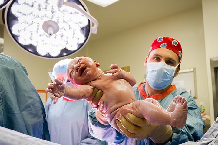
With its consistent growth, the smaller twin fetus was proving to be fatal to the older fetus by posing a danger of crushing the internal organs. That is when Dr. Miguel Parra-Saavedra, Monica’s specialist obstetrician took the decision to perform an emergency C-section to save the baby. On February 22, the seven-pound baby girl was delivered safely by the team via C-section to prevent further damage to her internal organs. The girl was named Itzamara. The next day the doctors performed a critical keyhole surgery on the little Itzamara to remove the fetus. Following removal, the fetus measured 45 mm in length and weighed 14 grams. However, the fetal brain and heart had not developed although the arms and legs had. It died the moment the little umbilical cord was cut (2).
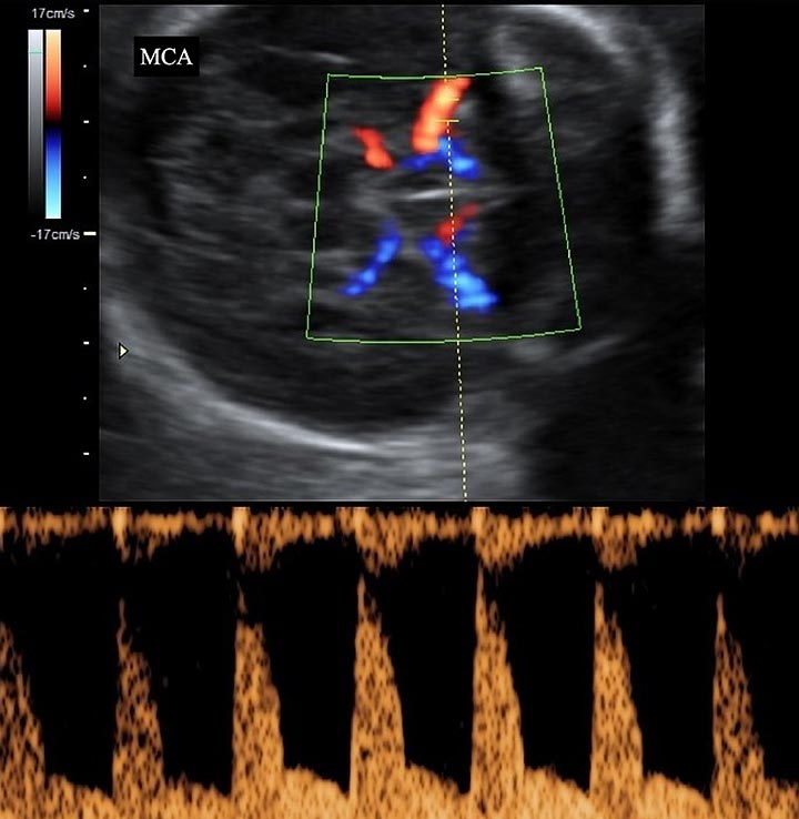
It has now been a month since Itzamara’s birth and she has been doing well with no signs of internal damage to her organs or stomach so far. According to Dr. Parra-Saavedra, it was one of the fascinating and strange cases in maternal-fetal medicine, probably of his career too. Monica’s case was referred to him by her primary obstetrician after she spotted the cyst on the fetus’ liver. The advancement in medical technology also played a significant role in the treatment of Itzamara. The doctors had used Color Doppler, which creates a multi-colored, detailed image of the blood vessels for closer inspection. It was due to this image that the doctor was able to conclude that what seemed like a cyst initially was, in fact, a tiny fetus growing inside the primary fetus, with its own umbilical cord.
While Itzamara’s condition did shock Dr. Parra-Saavedra, it was her mother who was incredulous. She could not believe that something like this could be possible. However, after the doctor explained to her each thing step by step, she eventually understood. It might have indeed been a very complicated surgery considering the age of the baby, but Dr. Parra-Saavedra is only too happy that Itzamara is doing well. Except for a slight scar, she seems to be hale and hearty, blissfully unaware that the whole world is talking about her!


